This week’s BEACON Researchers at Work blog post is by MSU graduate student Kyle Card.
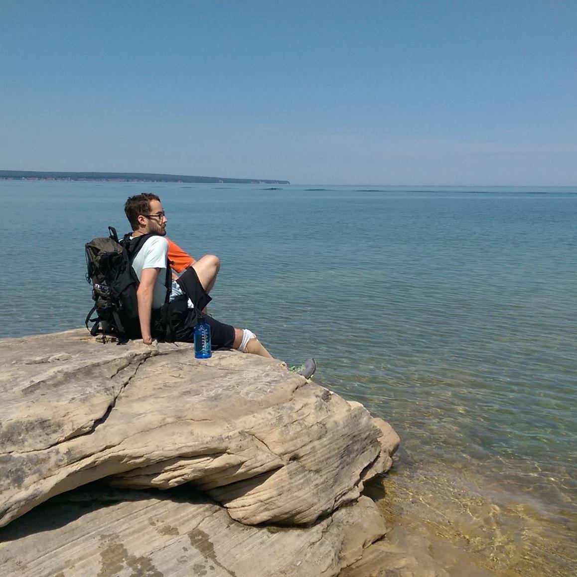
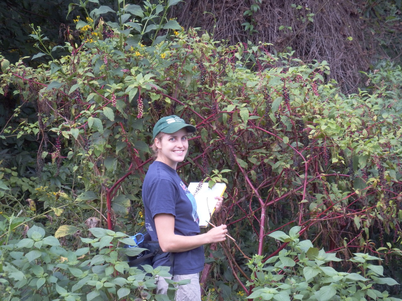
I visited the Pictured Rocks National Lakeshore over this past summer. Here I am sitting near Miners Castle looking toward Lake Superior.
Richard Feynman was an eccentric theoretical physicist and Nobel laureate who had a profound impact on the field of quantum mechanics. As a child, he would take long weekend walks with his father in the woods. During these walks, his father taught him about the workings of nature and the importance of observation. He learned that it was all right not to know the answer to something. Rather, the journey to find the answer is fulfilling and exciting in its own right.
A classmate, who knew of these walks, approached Feynman in the schoolyard one day and pointed to a bird sitting on a distant fence. He asked smugly, “What is the name of that bird?” After Feynman’s puzzled silence the boy chided in, “It’s a brown throated thrush. Your father doesn’t teach you anything!”
Feynman would tell this story to stress a lesson his father had taught him: it doesn’t matter what the bird is called. One can learn the name of a bird in all the languages of the world and still know absolutely nothing about the bird. There is a fundamental difference between knowing the name of something, and knowing something. The observation of the bird’s behavior is what counts. Science is about coming to a deeper understanding of nature. We are constantly pushing outward the boundary between what is known and what isn’t, which often leads to more unanswered questions to explore. This has led some to liken nature to an onion: when its layers are peeled back, one often uncovers more layers. Or as Philippe Verdoux put it, “Enlightenment leads to benightedness; Science entails nescience.” Ultimately though, the scientist takes pleasure in the simple act of discovery itself.
Like Feynman, I have always been deeply curious about nature. However, for most of my childhood I was fascinated by the cosmos. At the age of seven I was reading about astronomy and trying to wrap my head around nuclear fusion. By the age of thirteen I awaited the release of Stephen Hawking’s book The Universe in a Nutshell like it was the next Harry Potter installment (I also grew up reading the Harry Potter series, so I was a somewhat normal child, I swear!). I remember the librarian remarking how the book may be too advanced for me and I should consider looking for another book. I didn’t, and well, she was right – sort of. It was advanced, but why should I have let that deter me? It wasn’t until high school that I became interested in biology, and not until half way through my undergraduate studies that I became fascinated with the living world that can only be seen through a microscope.
I am a microbiologist who learned about evolution on my own to better understand my undergraduate research. I find evolution beautiful and elegant, and I am often taken aback by its immense explanatory power. I have become particularly interested in how genes interact with other genes in the same genetic background, a phenomenon called epistasis, and how chance historical events can constrain evolutionary outcomes. To introduce these, I invite the reader to imagine the game Plinko from The Price is Right.
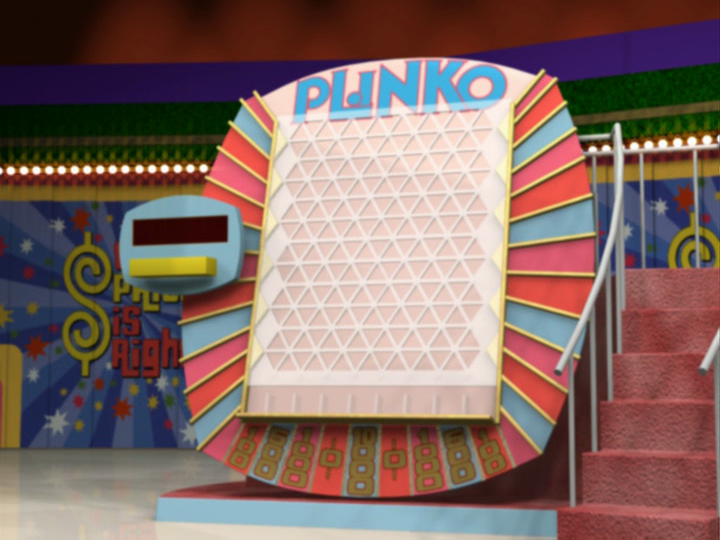
The Plinko board, in all its glory.
For those unfamiliar with the game: the Plinko board is very large and sits upright at a slight angle. Many small pegs protrude from its surface and at the bottom of the board lies nine containers. Each container has a corresponding prize amount. A lucky contestant climbs to the top of the board where he or she drops chips, one at a time, onto the pegs underneath. As the chip falls down the board it strikes the pegs, one after another, until it lands into one of the containers at the bottom. Whichever container the chip lands in, the contestant wins that amount. The best outcome for the player is to have the chips fall into the center container, which has the highest payout. The path a chip takes to the bottom varies every time the game is played. When the chip hits a peg, it may go left or right. The next peg it hits is dependent upon the direction it took during the preceding event. This process occurs all the way down the board until the chip lands in a container. If one were to imagine this occurring many times, each time a chip is released, it will take a slightly different path toward the bottom, and which container it falls into will likely differ between games. Thus, the outcome (reward) is dependent upon all preceding events that came before, along with the initial starting position of the chip itself. The outcome is further constrained by the limited number of containers; “the paths are many, but the destinations are few,” according to Simon Conway Morris.
Stephen Jay Gould proposed that a similar path dependence, which he termed historical contingency, largely makes evolution unpredictable. If one were to “rewind and replay the tape of life” multiple times, different evolutionary outcomes will emerge based on the sequence of historical chance events that occur. Gould’s proposal has been controversial, and only empirical investigation of it will determine if it is correct.
Historical contingency may play an important role in antibiotic resistance. Antibiotic resistance is a serious and pervasive threat to healthcare. According to the CDC, two million people will acquire antibiotic resistant infections every year in the United States alone. Among those two million, 20,000 will die. Antibiotic resistance is a product of natural selection in pathogen populations, and therefore, it is important to study this process from an evolutionary perspective. History may be important, because when a resistance mutation is introduced into a given genetic background, either through chromosomal mutation or on a plasmid, the level of resistance and fitness cost of the mutation are determined in part by the mutation’s interactions with other genes in that background. And the background itself is the product of a long history of chance evolutionary events, much like the Plinko example above. I am studying historical contingency’s role in antibiotic resistance using the long-term evolution experiment (LTEE) with Escherichia coli in the lab of Dr. Richard Lenski.
The LTEE consists of twelve E. coli populations that were founded from a single common ancestor and have been evolving for 63,500 generations, or over 27 years. Thus, it offers a unique opportunity to study the impact of history on evolution, as has been demonstrated by the work done on the evolution of aerobic citrate usage in one population. In the case of antibiotic resistance, the same resistance mutation may have different levels and costs of resistance in different LTEE populations because they have different genetic backgrounds due to their different evolutionary histories during the experiment.

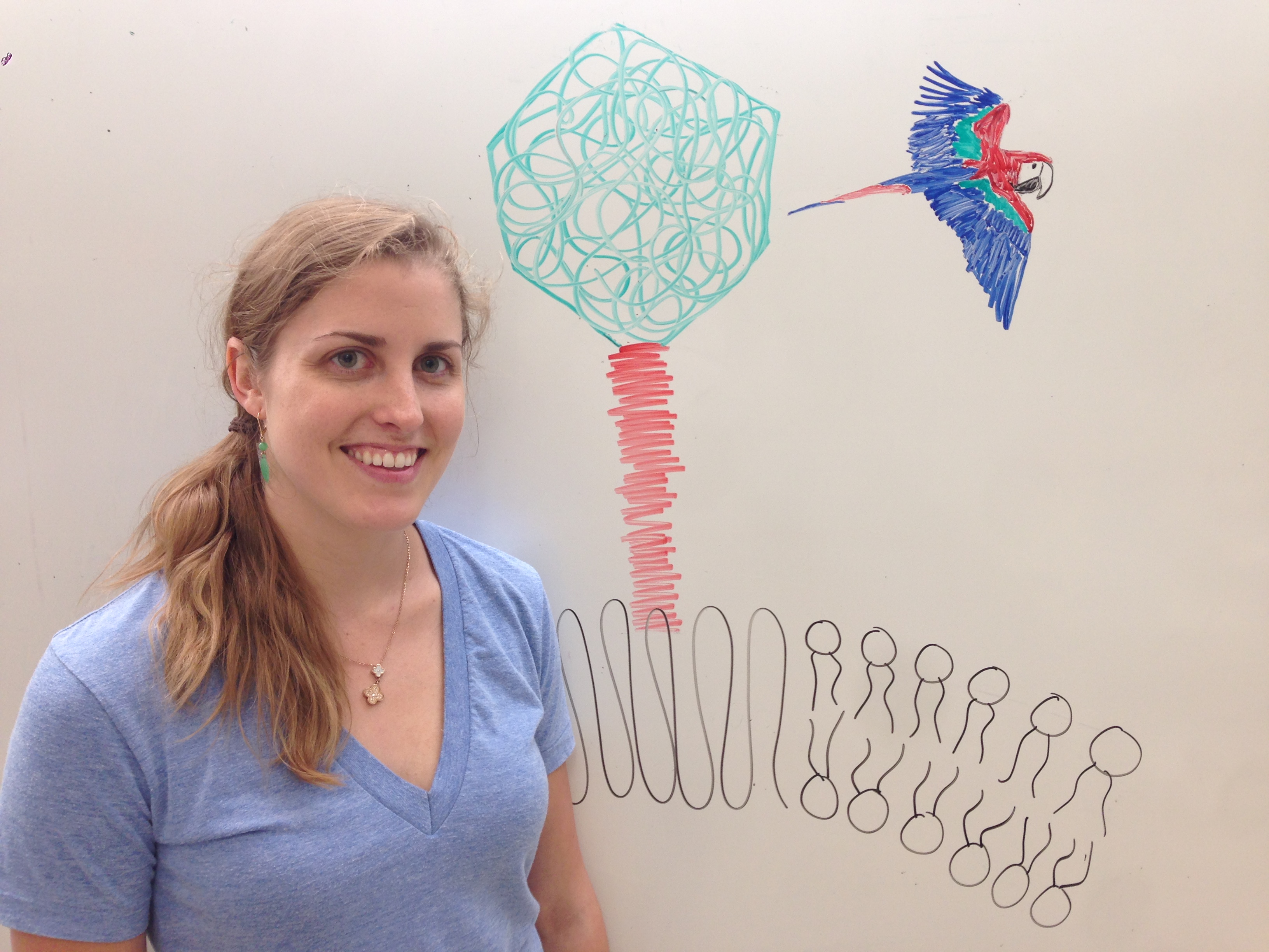
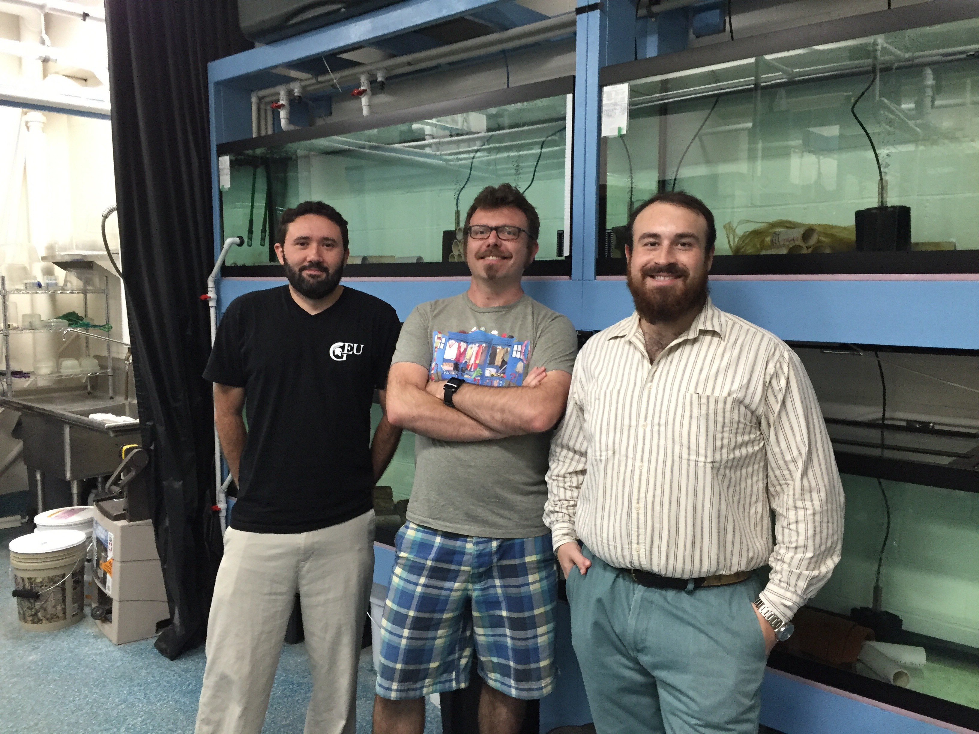
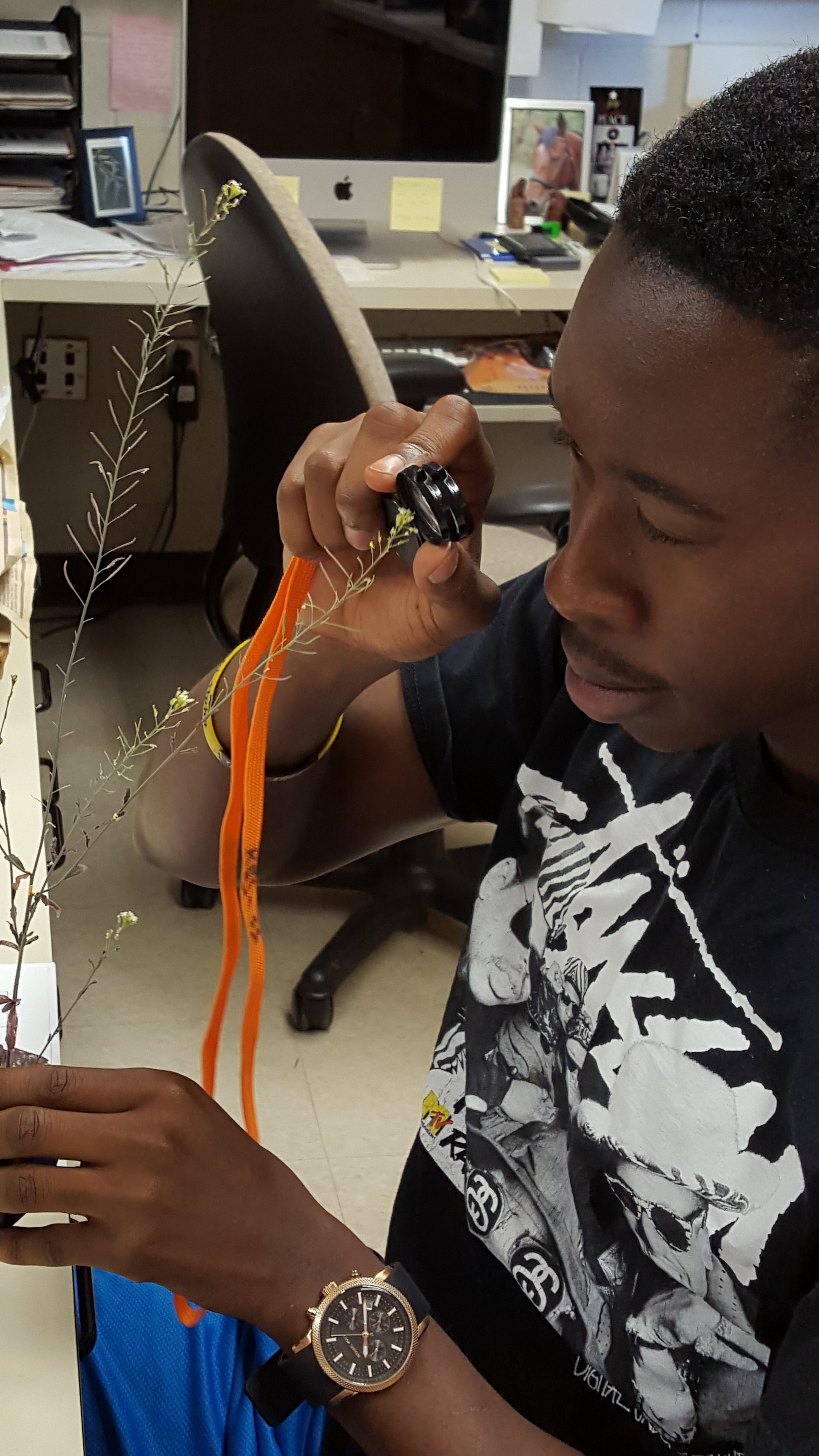
Look mom, I’m doing science!
As a new graduate student in the lab, I am still in experimental “dry dock.” In the coming months, I aim to test the level and cost of resistance of multiple antibiotic resistance mutations in the ancestor and clones isolated from all populations in the LTEE at 60,000 generations. I will test level of resistance using minimum inhibitory concentration assays. (This is just a fancy way of saying I will introduce increasing levels of antibiotics to my bacteria until they die. Then I will note how much antibiotic it took to kill them. I’m sorry little guys, but it’s for science!). To measure any fitness costs of resistance mutations, I will run pairwise competition assays comparing resistant mutants to their sensitive kin without the antibiotic present.
The observation of different levels and costs of resistance among multiple LTEE populations would suggest that historical contingency is likely playing a role in antibiotic resistance. Moreover, studying antibiotic resistance as an outcome dependent upon antecedent events is important to understanding its role in diverse medically relevant microbes with varied evolutionary histories.
Separate from this project’s medical relevance, I find the questions driving the research deeply interesting. For as long as I can remember I have been keenly fascinated with history. As a kid growing up, I watched the History channel quite a bit (before its sad downturn into Ancient Aliens and Pawn Stars), as well as The Price is Right (as any sick child staying home from school can attest). So maybe it was inevitable I would find a way to infuse both into my research, at least to some degree.
For more information about Kyle’s work, you can contact him at cardkyle at msu.edu