A microbe-dependent world: studying the legume-rhizobia symbiosis for a more sustainable future

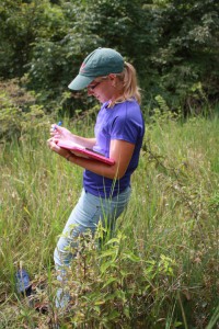
This post is by MSU grad student Shawna Rowe

Living in a world full of fascinating visual elements and intriguing macro-organisms often results in people forgetting the most abundant group of earth’s inhabitants— microbes. Microbes are not only the most abundant and diverse group of living organisms but are also, in my personal opinion, the most fascinating. Whether it be the Demodex brevis that colonize human faces or the rhizobia that live in our soils or the Thermus aquaticus that live in the depths of Yellowstone, microbes are inescapable and responsible for endless biological processes.

One group of bacteria, rhizobia, are soil-dwelling and underappreciated powerhouses of agricultural productivity. These bacteria form a specialized relationship with leguminous plants (soybean, bean, lentils, peanuts, etc.) in which they supply nitrogen, a globally limiting resource, in exchange for carbon. When undisturbed, this interaction naturally increases soil nitrogen content. Agricultural soils are frequently nitrogen limited which causes farmers to deposit approximately 80 million tons of nitrogen fertilizers on agricultural fields each year! This practice has resulted in increased crop yields at the expense of the environment. Toxic algal blooms pollute water sources, microbial communities have been destroyed, fossil fuels are burned to produce the fertilizers, and gaseous nitrogen compounds are released into the atmosphere as consequences of modern fertilizer production and use. Fortunately, the relationship between legumes and rhizobia offers an opportunity to offset the excessive use of fertilizers and begin shifting away from these environmentally detrimental practices.

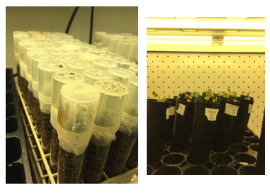
Medicago truncatula, a model legume on which I conduct research growing in two different types of growth containers. The fully encased one (test tubes) provides sterile conditions for assays that require a more controlled environment.

In this relationship the host legume provides the infrastructure in the form of specialized organs known as nodules. Inside these nodules live the hardworking rhizobia. The plant nodules serve as a protected space for the microbes to reproduce and expand as they complete the energy expensive task of converting N2 to NH3. Years of evolutionary pressure has resulted in a very tightly controlled balance of resource trade. However, as with most relationships there exists opportunities for trouble— in context of this mutually beneficial relationship the rhizobial partners have the opportunity to take more resources from the host plant while supplying comparably less nitrogen. This act has been termed “cheating.” Cheaters are problematic since they threaten to destabilize the long-established and important relationship; a reality that would further strengthen our dependence on nitrogen fertilizers in the agriculture sector. In Dr. Maren Friesen’s lab, I aim to elucidate molecular mechanisms of this resource trade between legumes and rhizobia. My work focuses on understanding how host plants are able to differentially recognize and respond to rhizobial partners of varying effectiveness. Developing an understanding of these response and control mechanisms is critical to understand how microbes are able to exploit their hosts and how external pressures are driving the emergence of cheaters.

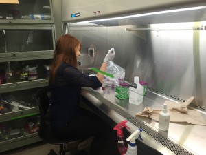
Shawna working in a biosafety cabinet in the Friesen lab space

As a native of southwest Missouri, ranked 6th in soybean production in the U.S., I spent most of my life surrounded by agricultural fields. Traveling to school frequently involved getting stuck behind a tractor when planting season arrived. Future Farmers of America was the largest student organization and roughly half of the student population had milked a cow before the age of 10. Although charming and hardworking, small agricultural towns are often times inherently (but unintentionally) anti-science. STEM education was severely lacking and evolution was a dirty word capable of eliciting dramatic arguments and endless frustration. Because of this, I loathed the idea of working in agriculture. Upon graduating high school, I entered college as a Biochemistry major with no clear idea of what “biochemistry” was nor what I could do with it. I was fortunate enough to land a job in a plant biochemistry research lab. There, they focused on understanding basic mechanisms of plant immune responses to pathogenic bacteria. That job set up the stage for my future research interests. I discovered the complex world of molecular signaling events and microbial associations. I learned about the co-evolution of organisms that commonly associate and how these associations drive the development and establishment of complex features of host-microbe interactions. I fell in love with the unseen world.

Years later, these experiences still serve as the foundation for the questions I ask and the topics I find intriguing. In the Friesen lab, I hope to better understand how hormones, specialized proteins, and various other plant derived molecules serve as regulatory components for the unique relationship leguminous plants have with the microbial world. Further developing our understanding of the regulatory mechanisms will both shed light on the co-evolution of legumes and rhizobia as well as the factors that threaten to destabilize this biologically important relationship.

Posted in BEACON Researchers at Work | Tagged , , , , | Leave a comment

Male battles split species apart

Picture of me: Behind me are some of the hundreds of fish tanks in the basement of Giltner containing all the baby sticklebacks we generated for this experiment.

This post is by MSU postdoc Jason Keagy

How do species form?

Stated more precisely, how does one species become two? This turns out to be an immensely difficult question to answer, because 1) species are not always distinct entities (species definitions are argued about ad naseum [1]) and 2) the formation of species (speciation) is a process that often takes a long time to complete.

One way in which species could form is if selection is divergent and a population responds to that selection [2] – for example, Anolis lizards that have adapted such that each species has limbs that are optimal for living in different types of vegetation [3], or insects that have specialized on feeding on different plants [4]. One way to represent the relationship between phenotypes (traits such as limbs, coloration, or digestive enzymes) and fitness is with a “fitness landscape” [5], so called because in three-dimensional representations (e.g., two traits as the x and y axes and fitness as the z axis), it can resemble a landscape of peaks and valleys. However, we don’t have a lot of great examples of these because it is often difficult to measure fitness and fitness often depends on multiple independent phenotypic traits in complicated ways.

The power of sticklebacks

In some freshwater lakes in British Columbia, you can find two different types of stickleback, called “benthics” and “limnetics” that are reproductively isolated, and therefore, typically referred to as species. These benthic and limnetic sticklebacks are descended from marine sticklebacks who bred in glacially fed streams. After the glaciers melted ~12,000 years ago, the weight of the ice being removed caused the land to rebound, and the uplifted streams became isolated lakes. Because of this relatively short timescale, these fish have become a model system for studying adaptation and speciation.

What is the difference between benthic and limnetic sticklebacks? Limnetics live in open water, eat plankton, and are more visually oriented, whereas benthics eat invertebrates off of plants or the lake bottom, live in complex spatially structured vegetated habitats, and are more dependent on smell. Limnetic and benthic sticklebacks also differ in body size, shape, and mating traits. In other words, they are really different! Critical for maintaining these differences is strong reproductive isolation and so the Boughman lab has long been interested in understanding what influences this isolation.

The role of male competition

Typically, the focus in speciation research has been on natural selection (even in sticklebacks). Much less studied and controversial is whether sexual selection can drive speciation. Especially unstudied is intrasexual (often male-male) competition’s role. That seems like a pretty big oversight to me. Flip on any nature show and you’re sure to see at least one scene of males bashing each other to pieces. It turns out Jenny Boughman, Liliana Lettieri, and I were already working on a project which was perfect for studying how male competition might impact speciation.

Fig. 1. Males compete intensely over territories on which they build nests. Pictured here are three males in a tank at KBS. The male in the foreground is directly over his nest. It’s pretty well concealed!

Male sticklebacks compete for territories on which they build their nests (Fig. 1). They’ll even destroy each others’ nests and steal pieces such as the choicest algae. Eventually, these males will try to attract females via courtship behavior to convince them to lay eggs in their nests. Male competition is extremely important to determining male fitness: if males can’t successfully obtain and keep a territory, and build and keep a nest, they are unable to reproduce (we rarely see sneak spawning). Male competition could have important impacts on speciation because males of each species build nests very close to each other in nature and are therefore direct competitors for space and resources.

Our research

Our main research questions included: How do male phenotypes relate to male competitive fitness? Do the resulting fitness landscapes have multiple peaks? Would these peaks promote speciation? We created hundreds of hybrid males in the laboratory through artificial crosses. This greatly expanded the combinations of phenotypes from that seen in the wild. Then we put these males in large outdoor tanks at Kellogg Biological Station that had sand and algae and food caught from nearby ponds. We measured lots of physical traits on the males and spent hundreds of hours recording their male competition behavior (with the help of an awesome army of undergrads).

Fig 2. Be really careful about what you are taking with you into water bodies. Your actions can have serious evolutionary and ecological consequences!

Our research revealed some surprises [6]. First, there were indeed two fitness peaks corresponding to pure benthic and pure limnetic multivariate phenotypes. But there was another region of high fitness (a bridge connecting the peaks) that implies certain intermediate hybrids were also good competitors. Interestingly, these hybrids had phenotypes like fish now seen in Enos Lake, where after anthropogenic disturbance (someone released crayfish into the water, Fig. 2) formerly distinct benthic and limnetic species are now a hybrid swarm (a depressing example of evolution in action). Previously the hybridization had been attributed to the crayfish’s introduction resulting in generalist rather than specialist sticklebacks having higher survival, a change in natural selection [7]. These generalists would have been produced by hybridization, which before happened at inconsequential numbers, but this trickle would have become larger as hybrids were now surviving to adulthood. However, our results show that sexual selection through male competition may also have been a contributing factor that sped up the species collapse. The hybrid males with phenotypes corresponding to the bridge within our fitness landscape would have likely been very successful at getting nests, increasing the likelihood of further hybridization. Our data strongly suggest male competition could be very important in the speciation process and impact speciation in complex ways.

Notes
[1] As one example of this disagreement, see Wu, C-I. 2001. The genic view of the process of speciation. Journal of Evolutionary Biology. 14: 851-865 and the ten responses.
[2] For a book dedicated to this topic, see Nosil, P. 2012. Ecological speciation. Oxford: Oxford University Press.
[3] A nice HHMI video description of this research is here: http://www.hhmi.org/biointeractive/origin-species-lizards-evolutionary-tree
[4] There are many nice examples of this including 1) pea aphids that have diverged to specialize on red clover and alfalfa, 2) fruit flies feeding on different species of cactus, 3) the races of apple maggot fly that feed on either hawthorn or apples, and 4) stick insects adapted to wildly different plants in California.
[5] There is some disagreement over the what specifically “fitness landscape” refers to and what the proper term is for what I refer to as a “fitness landscape” here (especially among philosophers of science). You can read about it in the first section of this book: Svensson, E., Calsbeek, R. (eds) 2012. The Adaptive Landscape in Evolutionary Biology. Oxford: Oxford University Press.
[6] Keagy, J., Lettieri, L., Boughman, J.W. 2016. Male competition fitness landscapes predict both forward and reverse speciation. Ecology letters. 19: 71-80.
[7] Behm, J.E., Ives, A.R., Boughman, J.W. 2010. Breakdown in postmating isolation and the collapse of a species pair through hybridization. American Naturalist. 175: 11–26.

Posted in BEACON Researchers at Work, Notes from the Field | Tagged , , , , , , | Leave a comment

Can evolution help us rebuild native habitats?

This post is by MSU graduate student Anna Groves.

If you look at the lyrics of two of the most iconic songs in American history, you’ll find that both reveal the composers’ fondness for the wide open spaces of our American landscape.

♪ Oh give me a home, where the buffalo roam… and the skies are not cloudy all day. ♫
Home on the Range, 1870s

♪ Oh beautiful, for spacious skies, for amber waves of grain… ♫
America the Beautiful, 1893

The concepts are really quite similar. But now that I study prairies as a graduate student at Michigan State University, I can’t help but notice the clear difference between the earlier and later song. What was once fondness for natural prairies becomes fondness for something that sounds a lot like a wheat field. Of course, you could perhaps cite the origins of the composers (one a pioneer, one an easterner visiting the west) or probably numerous other reasons why the lyrics are different, but to me, this is a clear illustration of what was happening across America around that time—we plowed up our grasslands and planted crops, and without thinking twice, we kept on plowing until there was almost nothing left of the native ecosystem.

Historical North American tallgrass prairie range in dark green; estimated remaining range in red. Prairie species pictured: American bison (Bison bison), Greater Prairie Chicken (Tympanuchus cupido), Regal fritillary (Speyeria idalia), and Bobolink (Dolichonyx oryzivorus).

Humans have converted a quarter of Earth’s land area for our own use [1]. Some habitats and areas have been more affected than others— nearly half of temperate grasslands have been lost globally, and in some places in the U.S., this number approaches 99.9% [2]. That’s a near complete loss of ecosystems like our own North American tallgrass prairie.

Luckily, not all converted land is still in use, and there are lots of opportunities to restore native systems back into the landscape. Ecological restoration is the practice of rebuilding or rejuvenating native ecosystems that have been degraded or destroyed by humans.

It’s easy to think about the ecology of native systems—especially restored systems— with some nearsightedness. If we re-plant a native prairie tomorrow, the goal is to make it work like a fully-functioning tallgrass prairie ASAP. There are businesses that will install a prairie on your property for you, and only the most patient and ecologically-savvy customers would accept “it’s mostly European weeds now, but give it 100 years, it’ll be great!” as a business model. Indeed, there is urgency from a conservation standpoint, too—at current rates of destruction, we need to start rebuilding for global stability of the next few decades, not centuries. There’s just no time to think about things like evolution—which happens over millions of years—is there?

Yours truly, pre-graduate school, on the edge of a remnant prairie somewhere in western Illinois. Photo by Randy Nyboer.

In reality, when we try to restore systems, we are likely not looking forward—or backward—far enough. Much of tallgrass prairie restoration is focused on the plant community, since the plants make up the structural habitat that all the other mammals, insects, and such live in. As it turns out, there’s a lot we can learn from thinking about the evolutionary histories of the plants we’re trying to restore.

This idea has been on my mind more than usual since last weekend, when I attended the Society for Ecological Restoration’s Midwest-Great Lakes annual chapter meeting. This group really knows their prairies, and are not only the best and brightest in their area, they are probably some of the best and brightest globally when it comes to restoring native grasslands. The keynote speaker [3] was Doug Ladd, the Missouri director of conservation for The Nature Conservancy. He has been a champion for tallgrass prairies for decades, and literally wrote the book on tallgrass prairie wildflowers (it’s called Tallgrass Prairie Wildflowers).

Last weekend, Doug reminded us to remember the evolutionary history of our plants. To remember that the first grasses came into being 55 million years ago. Angiosperms (flowering plants) came after, and the first oaks (the quintessential prairie tree) appeared 35 million years ago. Although our plants are ancient, so much of the North American prairie landscape was covered with a giant glacial ice sheet until just 15,000 years ago. If you’re a 350-year-old oak tree, that’s less than 50 generations. And for nearly all of the time that’s passed since the glaciers melted, human beings lived in the Midwest as well—the Native Americans. Only very, very recently in evolutionary history did Europeans settle the U.S., finally bringing their plows and oxen [4] to replace the prairies with their beloved amber waves of grain.

So, what does this tell us about tallgrass prairie restoration? It can be discomforting to think about the possibility that none of our ancient North American species actually had time to adapt to the post-glacial Midwestern landscape—especially now that we’ve gone and torn it all up, altered the soil chemistry and structure, and introduced countless other species from other continents to the landscape. The tallgrass prairie must be doomed, right?

My (current) favorite prairie picture. “Stiff sunflowers (Helianthus pauciflorus) greeting the sunrise in sand prairie. TNC Platte River Prairies, Nebraska.” Prairie and photo by Chris Helzer, The Nature Conservancy. For everything prairie restoration (and more pretty pictures), check out his blog.

Wrong! The plants are adapted, and we have learned so much from looking to the past and considering the adaptations of our species. In fact, knowing which species are adapted to which types of sites (e.g. dry versus wet) is Restoration 101! And more and more, people are finding evidence that it’s important to use local ecotypes in restoration—which means collecting seeds from a plant population as close to the restoration as possible—so that plants are as adapted to local conditions as possible [5]. Finally, we know that prairies are incredibly fire-dependent systems, and adapted in a landscape full of bison and other large grazers, as well as Native Americans who lit fires in their hunts. Some of our absolute best restoration success stories have come from large restoration landscapes that have re-instated fire and grazing management—some even re-introducing bison— that reflects what a prairie would have experienced once upon a time.

Restoration ecology (the science behind restoration) is continually finding ways to utilize plant adaptations to improve how we restore. A popular line of research today is to use plant traits—qualities like deeper roots, bigger leaves, and height of plants—to match up species with the landscapes they are best adapted to. Another is to consider how the history of a particular site, in terms of which species established when, will influence which plants will persist at that site. These are just two of the many ways in which ecology is informed by what we know about plant adaptions from considering the evolutionary history of our species. We may not think about “evolution” in those terms, but thinking about species and their interactions in this way is critical for our success in restoring prairies to the landscape.

I can’t promise skies that are not cloudy all day, but it seems increasingly likely we’ll once again be able to sing, oh give me a home, where the buffalo roam… ♫ all thanks to what we know about evolution.

http://www.annagroves.com
Follow me on Twitter: @annamgroves

[1] Hoekstra et al. 2005 Ecology Letters 8:23-29.
[2] Sampson & Knopf 1994 Bioscience 44(6):418-421.
[3] You can listen to Doug Ladd’s SER-MWGL keynote speech.
[4] Oh, my goodness. Did you know that the ox isn’t a unique species? It’s just a regular cow—usually a castrated male—that’s been trained as a draft animal. This city girl learned that fun fact embarrassingly recently. Last Monday.
[5] McKay et al. 2005 Restoration Ecology 13(3):432-440.

Posted in BEACON Researchers at Work | Tagged , , , , | Leave a comment

UI BEACON Director James Foster to Receive Highest Faculty Rank at the University of Idaho

This post is by UI IBEST Communications Coordinator Amberly Beckman.

University of Idaho Distinguished Professor James A. Foster

The University of Idaho recently named Prof. James A. Foster a University Distinguished Professor, the highest rank for faculty at the University. Foster is one of only fourteen faculty members to receive this honor, since its inception in 2011.

The University of Idaho established the rank of Distinguished Professor to acknowledge faculty who embody excellence in teaching, scholarship, outreach and service. The selected nominees are faculty who have been with the University for at least seven years, and received national or international recognition.

Foster is a professor of Biological Sciences with a background in evolutionary biology, computer sciences, and philosophy. His work with both the Institute for Bioinformatics and Evolutionary Studies, and the Beacon Center for the Study of Evolution in Action has brought together interdisciplinary study of evolution across campus and the country.

He founded the IBEST Computational Resources Core, co-founded the Bioinformatics and Computation Biology program at IBEST (with Prof. Paul Joyce), and co-founded IBEST itself (with Prof. Holly Wichman).  In addition, he is the director of the BEACON project at University of Idaho.

The University Awards for Excellence are held annually and recognize excellence at many academic levels. The ceremony is scheduled for 6:00pm on Tuesday, April 16 at the Bruce M. Pitman Center’s International Ballroom.

Also receiving the rank of University Distinguished Professors are Paul Joyce, and Nilsa Bosque-Perez. Past IBEST awardees include Holly Wichman, Larry Forney, and Lizette Waits.

Posted in BEACON in the News, BEACON Researchers at Work, BEACONites, Member Announcements | Tagged , | Leave a comment

BEACON in the News: (How) does a bear walk in the woods?

Researcher Katie Shine sets up equipment for a study of black bear locomotion at the Oregon Zoo. (Photo by Michael Durham, courtesy of the Oregon Zoo)

This week BEACON grad student Katie Shine and her PhD advisor Craig McGowan at the University of Idaho are getting a lot of news coverage for their research investigating bear locomotion using live bears from Washington State University (grizzlies) and the Oregon Zoo (black bears)!

Oregon Zoo black bears help out with walking study
“The Oregon Zoo’s black bears Cubby and Takoda recently offered up their paws for the sake of science.”
The Oregonian/April 4/Laura Frazier

The Bear Necessities Of Walking
“Flat-foots are far less studied than their tiptoe counterparts, and scientists … recently traveled to the Oregon Zoo, where keepers helped them lead black bears Takoda and Cubby through the same walking trial.”
1190 KEX News Radio/March 30
This article was originally on Fox12Oregon KPTV and picked up by other Fox affiliates around the country, including Fox 5 Vegas; KITV (Honolulu); KBZK (Bozeman, MT); KRTV (Great Falls, MT); and KXLH (Helena, MT).


Posted in BEACON in the News, BEACON Researchers at Work, Notes from the Field | Tagged , , | Leave a comment

Introducing BEACON’s New Science Outreach and Communication Postdocs

Alexa Warwick out in the field at night with her study organism, the Pine Barrens Treefrog.

This post is by MSU postdoc Alexa Warwick.

Alexa Warwick is the new Evolution Education and Outreach Postdoc at BEACON working with Dr. Louise Mead.

Alexa worked on her Ph.D. with Drs. Emily Lemmon and Joseph Travis at Florida State University focusing on the ecology, evolution, and conservation of the Pine Barrens Treefrog (Hyla andersonii; Fig. 1). At BEACON, she will be coordinating three established outreach programs that were previously run by Dr. Jory Weintraub. These annual programs include the Darwin Day Roadshow, Ecology and Evolution events at the National SACNAS Conference, and the Undergraduate Diversity Travel Award to attend the Evolution meetings (this year in Austin, TX; award applications due April 18th!). The programs are collectively sponsored by BEACON, the Society for the Study of Evolution, the Society of Systematic Biologists, and the American Society of Naturalists.

Fig 1. The Pine Barrens Treefrog has a unique species distribution across three isolated regions in the eastern United States.

In addition to these national outreach programs, Alexa will also be involved with developing and assessing other outreach/education projects. One of these projects is an efficacy study of Data Nuggets in high school biology classes. Data Nuggets are an exciting way to introduce K-16 students to real scientists and authentic research data, while also helping scientists increase their broader impacts. Alexa is passionate about effectively engaging students and the general public with science research, so feel free to contact her with other ideas for outreach/education collaborations!

She will also be continuing to do research on amphibians while at BEACON. For example, she is working with Dr. Elise Zipkin’s lab at MSU and SPARC-net, a regional group working to understand climate and land use change on salamander population dynamics. For this project salamander sampling plots will be established at the Kellogg Biological Station and the data collected there will be combined with similar plots across the northeastern United States.

A gladiator frog (Hypsiboas pugnax) that Alexa caught in the lowlands of Colombia.

Alexa’s other research interests include studying the evolutionary history of organisms using genomic data (phylogeography/phylogenetics) and the effects of behavioral interactions on the evolution of species (sexual selection). Her work on the Pine Barrens Treefrog has addressed the origin and maintenance of its unique distribution (Fig. 1), and she has recently started working with gladiator frogs (genus: Hypsiboas) in South America. This is a fascinating group of frogs with rather unusual behaviors; they actually get their common name from the male-male combats that occur using a spine on their ‘thumbs’. Check out Alexa’s website for more details about current research projects or send her an email at awarwick [at] msu.edu.

Also check out our other two previous New Science Outreach and Communication Postdocs posts by Wendy Smythe and Travis Hagey

Posted in BEACON Researchers at Work, BEACONites, Notes from the Field | Tagged , , , , | Leave a comment

A Mighty Mouse and a Scientist in Training: A Story of Physiological and Personal Evolution

This post is by Okemos High School student Maddie Stover working in Dr. Ashlee Rowe’s lab in the Neuroscience Program & Department of Integrative Biology at MSU.

A particularly effective nervous system makes the four-inch grasshopper mouse quite a fearsome desert creature, especially if you are an arthropod. Not only do these rodents howl to defend their territory and stalk their prey like big cats, but they hunt venomous scorpions, centipedes, and tarantulas. Yet these arthropod preys are not without a strong defense; their venom contains potent neurotoxins that are designed to both incapacitate and induce intense pain in their predators. In an evolutionary arms race, grasshopper mice (Onychomys​ spp.), when compared to house mice, exhibit reduced sensitivity to the pain caused by bark scorpion (Centruroides​ spp.) venom (1). Our lab is investigating the molecular and physiological adaptations to the nervous system of grasshopper mice that confer resistance to bark scorpion venom. Three species of grasshopper mice are distributed throughout the deserts and prairies of North America; I study the Chihuahuan desert species, Mearns’ grasshopper mouse (O​. arenicola)​. Specifically, I am interested in the adaptations underlying their resistance to the venom of the striped bark scorpion (C​. vittatus)​.

A grasshopper mouse attacking a bark scorpion. Photo by Dr. Matthew Rowe.

Pain signals are transmitted to the central nervous system (CNS) by way of ion channels embedded in cell membranes. Sodium (Na+) and potassium (K+) ion channels play an important role in pain-signaling. As these channels are essential for many physiological processes they are very well conserved, such that a few amino acid substitutions can produce a significant change in the overall electrical activity of the channels. Bark scorpion venom contains low-molecular-weight proteins (toxins) that bind and manipulate ion channels, causing intense pain. Rowe et al. (2) previously showed that the Sonoran desert grasshopper mouse (O​. torridus)​ is resistant to the burning pain of its local prey, the Arizona bark scorpion (C​. sculpturatus)​, via amino acid changes to a pain-pathway Na+ channel (Nav1.8). Instead of inhibiting toxin binding to its target channel (Nav1.7), amino acid substitutions in O. torridus​ Nav1.8 enable the channel to bind a protein in C​. sculpturatus​ venom, ultimately blocking the pain signal the venom is trying to send to the CNS (2). However, O. arenicola a​ppear to be resistant to C. vittatus​ venom through an alternative mechanism. Since C. vittatus​ venom contains many K+ channel toxins, our lab is looking at the structure and function of O​. arenicola K​ + channels to determine whether changes in these ion channels confer reduced pain sensitivity to C. vittatus​ venom.

A potassium channel consisting of an alpha subunit (red) and a beta subunit (blue). Pongs, O. & Schwarz, J.R. (3).

In addition to providing an opportunity to study evolution at the molecular level, the grasshopper mouse model might also have interesting implications for chronic pain. Currently, opioids are the most common medication used to treat chronic pain. However, these drugs can have unpleasant side effects and become addictive. In order to develop better treatments for chronic pain, a more detailed understanding of the ion channels involved in pain-signaling is required. In the grasshopper mouse model we are able to study the physiological effects of structural variation in different types of potassium channels that regulate pain-signaling.

When I first came to the Rowe Lab I was interested in how the structure and function of K+ channels contributed to the grasshopper mouse’s decreased sensitivity to toxin-induced pain. As I continued reading about these channels I found that K+ channels (the main pore region of which is called the alpha subunit) assemble with various auxiliary subunits. Beta subunits (a type of auxiliary unit) are cytoplasmic proteins that help regulate the channel’s electrical activity (3). Beta subunits are also the targets of certain scorpion toxins. At the Rowe Lab, I am studying whether K+ channel beta subunits contribute to the grasshopper mouse’s resistance to scorpion toxins.

To investigate this question I sequenced two genes that encode K+ channel beta subunits from O. arenicola.​ I also cloned these genes so that we may coexpress K+ channel alpha and beta subunits in a heterologous system, and record the electrical activity of this channel complex. I found structural variation, when compared to house mouse orthologs, in both beta subunits I am studying. Currently, we are testing whether these structural changes have functional consequences for the K+ channel that contribute to grasshopper mouse venom resistance.

BEACON-Maddie workingI have had a great first experience with research at the Rowe Lab. All of our lab members have been important mentors to me and have taught me so much. My lab helped me transition from a high school setting to a professional one (their patience, god bless) where I could learn skills that will be helpful in any area of work. I have learned that problem solving really is as big of a deal as they make of it on college websites and to never be ashamed to ask for help. I have learned that you really do regret what you do not write down, that white boards are the most important piece of equipment, and that I can really freak my grad students out by talking about my birth year.

As previously stated beta subunits are small (the gene is around 2,000 base pairs long) when compared to alpha subunits (the gene is ~8,000 base pairs long). They are also quite new to molecular biology, as they were discovered in 1992. I suppose I am analogous to a beta subunit in that we are both relatively small and new to science.

References

  1. Rowe, A.H. & Rowe, M.P., 2008. Physiological resistance of grasshopper mice (Onychomys spp.) to Arizona bark scorpion (Centruroides exilicauda) venom. T​ oxicon,​ 52(5), pp.597–605.
  2. Rowe, A.H. et al., 2013. Voltage-gated sodium channel in grasshopper mice defends against bark scorpion toxin. Science,​ 342(6157), pp.441–446.
  3. Pongs, O. & Schwarz, J.R., 2010. Ancillary Subunits Associated With Voltage–Dependent K+ Channels. P​ hysiological Reviews,​ 90, pp.755–764.
Posted in BEACON Researchers at Work, Education | Tagged , , , , , | Leave a comment

Deputy Director Charles Ofria Receives College of Engineering Withrow Distinguished Scholar Senior Award

Left to right: Erik Goodman (BEACON Director), Charles Ofria (BEACON Deputy Director), Leo Kempel (MSU College of Engineering Dean)

BEACON is very excited to congratulate our Deputy Director Charles Ofria on his selection for the College of Engineering Withrow Distinguished Scholar Senior Award. This is the MSU College of Engineering’s highest award for research and a significant achievement. The award is presented annually to one senior faculty member. A committee selects awardees after receiving nominations from all departments within the College of Engineering. In introducing Prof. Ofria for presentation of the award, BEACON Director Erik Goodman reviewed many of Prof. Ofria’s professional accomplishments, and noted especially his key role in generating the BEACON concept that led to its funding. Prof. Ofria continues to play a large role as Deputy Director and a leader of many projects. As Prof. Goodman stated, “Without Charles, there would be no BEACON.”

Posted in BEACON Researchers at Work, BEACONites, Member Announcements | Tagged , , | Leave a comment

A niche in time: adaptations in sensory processing associated with temporal niche

Andrea Morrow

MSU graduate student Andrea Morrow

This post is by MSU graduate student Andrea Morrow.

People often associate certain physical adaptations with an animal’s temporal niche, i.e. daily activity pattern. For example, some nocturnal animals have larger eyes, larger corneas, or higher concentrations of rod cells than diurnal animals [1]. These traits are valuable for animals that are awake and active at night because they augment the collection and utilization of what little light is available, which is advantageous when it comes to avoiding predators and finding resources. While these adaptations may be valuable, nervous tissue involved in processing sensory information requires a large amount of energy to develop, maintain, and power [2]. For this reason, it might not be a great idea for a nocturnal animal to invest extensive amounts of energy into processing complex visual information, when the amount of information obtained will always be restricted due to lack of light. Let’s think of the flip side to that. Would a diurnal animal, with plenty of light available, profit more than a nocturnal animal from expending substantial energy in visual processing?

In general, if the energetic cost is high, then the reward should also be high, or else the trait will not be maintained [3]. We can apply this concept of costs and benefits to all sensory processing. Brain size is constrained by energetic costs [4], and therefore we expect energetic trade-offs when it comes to investments in specific areas or tasks. How do nocturnal vs diurnal animals utilize visual, tactile, olfactory, and auditory cues from their environment? Are some more heavily relied upon by diurnal animals, and less by nocturnal animals? If, as I suspect, diurnal animals invest more in visual processing than nocturnal animals do, does it come at a cost in the form of other sensory processing?

Dr. Barbara Lundrigan (Department of Integrative Biology), Dr. Laura Smale (Department of Psychology), and I are trying to answer some of these questions by examining multiple sensory processing structures within the brain. We want to determine whether investment trade-offs between olfactory, visual, and auditory processing occur following evolutionary shifts in activity pattern.

Focal group

Determining the effect of a single factor in an observational study can be a heady task, especially when it comes to complex brain structures. Ecological, behavioral, and phylogenetic factors all work in shaping brain morphology and functionality. We are sampling widely across the order of Rodentia (which includes more than 2,000 extant species) in order to include species that vary in life history, behavior, environment, and evolutionary history. Within this broad framework, we are sampling closely related species within multiple subfamilies where temporal niche transitions have occurred independently. For example, with our colleague Dr. Paul Meek, we are sampling Australian bush rats (nocturnal) and Australian swamp rats (diurnal) from the family Muridae. We are also sampling Southern flying squirrels (nocturnal) and North American red squirrels (diurnal) from the family Sciuridae. Both of these groups have independently experienced activity pattern shifts. We want to capture as many independent evolutionary shifts as possible, as well as including animals that exhibit activity patterns other than strict nocturnality and diurnality. Our working list currently includes about 30 different species from 10 subfamilies, representing several independent shifts. We have collected tissue samples from 13 species and over 80 individuals to-date.

Structures of interest

Two coronal sections of an Australian rat. The top image is stained with Cresyl Violet. The bottom image is a section stained with Acetylcholinesterase. The Medial Geniculate Nucleus (MGN) is outlined and shaded in blue and the Lateral Geniculate Nucleus (LGN) is outlined and shaded in black.

The goal is to quantify the investment in olfactory, visual, and auditory processing. For olfactory processing, we are measuring the weight of the olfactory bulbs, relative to the weight of the entire brain, in each individual. For visual and auditory processing, we are measuring the volume of the Lateral Geniculate Nucleus and specific layers of the Superior Colliculus, which are visual processing structures, and the Medial Geniculate Nucleus and the Inferior Colliculus, which function in auditory processing. We do this by cryosectioning the frozen, unfixed brain tissue of each animal. The tissue is stained using two types of stain (Cresyl Violet and Acetylcholinesterase), microphotographs of each tissue section are taken and uploaded to the computer, and then each structure is delineated. These histological methods allow us to measure the area of each structure on each section of brain tissue, which we then use to calculate volume.

Our objective is to better understand how animals utilize the different sensory cues in their environment, as well as how investment in the processing of that information changes with the evolution of different activity patterns. While we don’t have data on the visual and auditory structures yet, we have collected some data on the olfactory bulbs. While the data are preliminary and represent only 13 species, with 1-6 individuals each, differences in relative olfactory bulb size between nocturnal and diurnal species are already apparent. The nocturnal species possess larger olfactory bulbs, relative to brain size, than the diurnal species. In addition to adding more species, we plan to look more thoroughly at the olfactory system in relation to activity pattern in the future.

I’ve always been interested in how organisms adapt to their environments and how their morphology and physiology are shaped (or constrained) by their evolutionary history and ecology. It is intriguing to imagine one environment (spatially) being separated temporally simply by the rotation of the earth and the ensuing cycles of light. The temporal niche presents many unanswered questions. What changes occur when animals adapt to a new temporal niche? Are these changes consistent and predictable? What conditions permit or restrict temporal partitioning? Is competition a driving force of this type of niche shift? Must there be plasticity, or flexibility, in activity pattern present for such shifts to occur, and if so, at what level? I’m hoping that, in time, I will be able to answer at least a few of these questions.

References

[1] Kirk EC. 2006. Effects of activity pattern on eye size and orbital aperture size in primates. Journal of Human Evolution. 51:159-170.

[2] Mink JW, Blumenschine RJ, Adams DB. 1981. Ratio of central nervous system to body metabolism in vertebrates: its constancy and functional basis. American Physiological Society. 241:R203-R212.

[3] Niven JE, Laughlin SB. 2008. Energy limitations as a selective pressure on the evolution of sensory systems. Journal of Experimental Biology. 211:1792-1804.

[4] Barrickman NL, Lin MJ. 2010. Encephalization, expensive tissues, and energetics: An examination of the relative costs of brain size in Strepsirrhines. American Journal of Physical Anthropology. 143:579-590.

Posted in BEACON Researchers at Work | Tagged , , , , | Leave a comment

How lemur social networks shape microbial transmission

This post is by UT Austin graduate student Amanda Perofsky.

Amanda Perofsky holding a sedated Verraux’s sifaka in 2012. The Sifaka Research Project at Ankoatsifaka Research Station captures animals periodically to mark them with collars, monitor health, and collect genetic material.

Primates exhibit diverse ecological and behavioral patterns, ranging from solitary foragers to several hundred individuals, as in the multi-level societies of hamadryas baboons [1]. Many wild primates live in social groups that range from stable to highly fluid, and this has important implications for the transmission of viruses, bacteria, and parasites between individuals. Several studies have demonstrated that host contact patterns alter the course of disease spread in wildlife populations [e.g., 2–6]. However, the effects of social contacts on commensal and mutualistic microorganisms, such as the mammalian gut microbiome, are just beginning to be appreciated [e.g., 3–5].

As a PhD student in Dr. Lauren Ancel Meyers’ research group at UT-Austin, my research aims to better understand the impact of primate social behavior on susceptibility to intestinal bacteria, and to identify factors that shape the gut microbiomes of individuals, social groups, and populations. In particular, I study Verreaux’s sifaka (Propithecus verreauxi), an endangered lemur species, in close collaboration with Dr. Rebecca Lewis, a primatologist in the Department of Anthropology at UT. Verreaux’s sifaka, like all lemurs, are unique to Madagascar, an island country off the southeastern coast of Africa. Dr. Lewis has studied sifaka behavior for over twenty years and directs the Ankoatsifaka Research Station located in Kirindy Mitea National Park in southwestern Madagascar. To distinguish between individuals, Dr. Lewis has fitted the sifaka at her field site with nylon collars and tags. One adult female in each social group has a radio collar, which enables researchers to locate specific groups in the forest using a radio transmitter.

A sifaka social group sunbathing in Kirindy Mitea National Park, Madagascar.

Sifaka live in small, tight-knit social groups that feed, sleep, rest, and travel together. Physical contact that promotes bacteria transmission may occur when sifaka groom each other, co-feed in trees, and huddle together. Within a social group, some pairs spend more time grooming each other than others [10]. Because sifaka (like all lemurs) groom with their mouths and toothcombs, do close grooming partners exchange intestinal bacteria more frequently compared to sifaka that rarely interact? Additionally, some individuals groom everyone in their social group, whereas others have only one or two close relationships. Are these highly social sifaka exposed to a greater diversity of microbes?

An unmarked sifaka grooms an adult female with infant nursing.

To answer these questions, I examine both sifaka social network structure and bacterial communities in sifaka fecal samples. Based on the behavioral data recorded by Dr. Lewis and her field assistants, I construct social network models that represent potential microbe-transmitting social contacts that occur within and between social groups. Social network models provide an intuitive framework for visualizing a population as a set of individuals (or, “nodes”) connected by “edges” [11] that, in this context, represent pathways for bacteria transmission.

In 2012, I traveled to Madagascar to collect fecal samples from each sifaka in the Ankoatsifaka study population and will be returning this summer to collect more samples. Each morning, we use a radio transmitter to locate the social group we’ll be following for the day. Collecting the fecal samples can be exasperating at times, and I couldn’t have accomplished my 2012 field season without the generous help of my collaborator, Elvis Rakatomalala, a Malagasy PhD student at the University of Antananarivo. Even with two people, it can be difficult to follow a large social group when the lemurs are jumping away in every direction and defecating all at the same time in different places. The fecal pellets scatter as they hit branches and leaves on the way down to the ground. We then crawl on our hands and knees through scratchy thorns, looking for pellets the size of Tic Tacs in the leaf litter. Once we locate a sample, we record the GPS point, the date, and the sifaka’s identity, and then put the pellets in a tube with liquid that preserves the DNA. The rest of the research— DNA extraction, bacteria sequencing, and data analysis— takes place back at UT.

After describing my fieldwork as difficult at times, why would I travel across the world to collect lemur fecal samples? Although collecting lemur fecal samples isn’t always easy, I love working outside surrounded by the forest,, and it’s fascinating to observe animals that can’t be found anywhere else in the world. Being in the field and collecting the samples myself gives me a deeper intuition for sifaka social behavior and its potential epidemiological consequences, providing insights that wouldn’t occur to me if I were at my desk in Austin.

  1. Griffin, R. H. & Nunn, C. L. 2011 Community structure and the spread of infectious disease in primate social networks. Evolutionary Ecology 26, 779–800.
  2. Böhm, M., Palphramand, K. L., Newton-Cross, G., Hutchings, M. R. & White, P. C. L. 2008 Dynamic interactions among badgers: implications for sociality and disease transmission. Journal of Animal Ecology 77, 735–745.
  3. Hamede, R. K., Bashford, J., McCallum, H. & Jones, M. 2009 Contact networks in a wild Tasmanian devil (Sarcophilus harrisii) population: using social network analysis to reveal seasonal variability in social behaviour and its implications for transmission of devil facial tumour disease. Ecology letters 12, 1147–57.
  4. Clay, C. A., Lehmer, E. M., Previtali, A., St Jeor, S. & Dearing, M. D. 2009 Contact heterogeneity in deer mice: implications for Sin Nombre virus transmission. Proceedings of the Royal Society B-Biological Sciences 276, 1305–1312.
  5. Craft, M. E., Volz, E., Packer, C. & Meyers, L. A. 2011 Disease transmission in territorial populations: the small-world network of Serengeti lions. Journal of the Royal Society Interface 8, 776–86.
  6. Rushmore, J., Caillaud, D., Matamba, L., Stumpf, R. M., Borgatti, S. P. & Altizer, S. 2013 Social network analysis of wild chimpanzees provides insights for predicting infectious disease risk. The Journal of Animal Ecology , 976–986.
  7. Tung, J., Barreiro, L. B., Burns, M. B., Grenier, J.-C., Lynch, J., Grieneisen, L. E., Altmann, J., Alberts, S. C., Blekhman, R. & Archie, E. A. 2015 Social networks predict gut microbiome composition in wild baboons. eLife 4.
  8. Moeller, A., Foerster, S., Wilson, M., Pusey, A., Hahn, B. & Ochman, H. 2016 Social behavior shapes the chimpanzee pan-microbiome. Science Advances.
  9. Bull, C. M., Godfrey, S. S. & Gordon, D. M. 2012 Social networks and the spread of Salmonella in a sleepy lizard population. Molecular Ecology 21, 4386–4392.
  10. Lewis, R. J. 2010 Grooming patterns in Verreaux’s sifaka. American Journal of Primatology 72, 254–261.
  11. Newman, M. 2010 Networks: An Introduction. Oxford University Press.


Posted in BEACON Researchers at Work, Notes from the Field | Tagged , , , , | Leave a comment